NEW MINDRAY DP-10 ULTRASOUND
Description
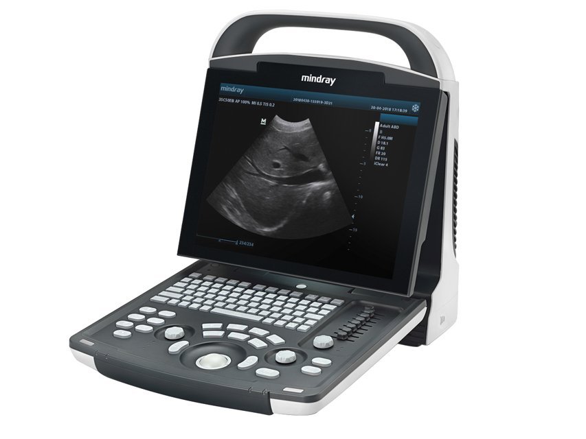
MINDRAY DP-10 DIGITAL ULTRASOUND SYSTEM
Good image quality, easy to use and lightweight to take it anywhere. it combines fashionable design, diagnostic confidence and economy.
• Display modes and imaging processing
Broadband, multi-frequency imaging: B, B/B, 4B, B/M, M
• Imaging processing
– multi-frequency probes for 2d imaging modes –
– iClear: speckle reduction image
– THi (Tissue Harmonic imaging) with convex probe 33976 only
– TSi (Tissue Specific imaging)
– zoom function: spot zoom, pan zoom
– ExFoV (Extended field of view)
Extended view of the anatomical structure on all convex and linear probes allow to discover better diagnostic information.
– iTouch (auto image optimization)
– colour map
• User interface
– alphanumeric QWERTY keyboard with backlight
– trackball: speed presettable
– 8-segment TGC
– user defined blank keys: shortcut for easy access to menus
• iStation™ intelligent workflow
Intelligent patient data management system
– integrated search engine for patient data
– detailed patient information review
– intelligent data backup/restore
– patient data/image sending, data deleting (USB , DVD, DICOM)
– exam managing: activate exam
• Storage/review mode:
– image archive on hard disk and USB mobile storage medium, temporary saving in cine memory, directly transfer report to PC
– 2 USB ports
– multiple image formats: BMP, JPG, dCM, FRM, aVi, dCM, CiN
– iVision: demo player
– Cine review: auto, manual (auto review segment can be set), supports linked cine review for 2d, M images
– Cine memory capacity (max.)
– clip length presettable: 1-60 s
B mode: 11959 frames
M mode: 110.0 s
Supplied with GB, iT manual and Cd manual (GB, FR, iT, ES, PT, dE, RU, BR).
• iScanhelper function
Dedicated inbuilt tutorial function to enhance ultrasound experience.
– wide selection of application specific exam planes
– anatomical illustrations
– standard ultrasound images
– scanning reference pictures
– tips on scanning and diagnosing
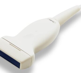
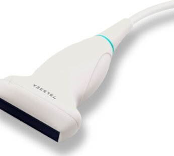
• Optional PW doppler for probes 33976 (Convex), 33977 (linear)
DP-10/20 support superb PW doppler imaging to access blood flow in different clinical conditions.
With PW, doctors will be able to:
– differentiate non echoarea and blood vessels
– distinguish arteries and veins
– evaluate blood flow velocity for more comprehensive diagnosis
– Check the health of a fetus by blood flow evaluation of the umbilical cordfeatures
Parameter adjustment: Gain, Sample volume size, Scale, Steer, dynamic range, Wall filter, etc.
Additional information
| Weight | 9 kg |
|---|---|
| Dimensions | 34 × 50 × 50 cm |
| Technical Specification | • Dimension: 16 x 29 x h 35.4 cm (6.3"" x 11.4"" x h 13.9"") • Weight: 5.1 kg • Display: 12.1"" LED, 50-60 Hz, CZ, DE, ES, high-resolution 1024 x 768 • Display language: FR, IT, PL • Power: AC 100-240V, PT, RU |


4 reviews for NEW MINDRAY DP-10 ULTRASOUND
There are no reviews yet.